The BMI lab bridges research in Machine Learning and Sequence Analysis methodology research and its application to biomedical problems. We collaborate with biologists and clinicians to develop real-world solutions.
We work on research questions and foundational challenges in storing, analysing, and searching extensive heterogeneous and temporal data, especially in the biomedical domain. Our lab members address technical and non-technical research questions in collaboration with biologists and clinicians. At the research group’s core is an active knowledge exchange in both directions between the methods and the application-driven researchers.
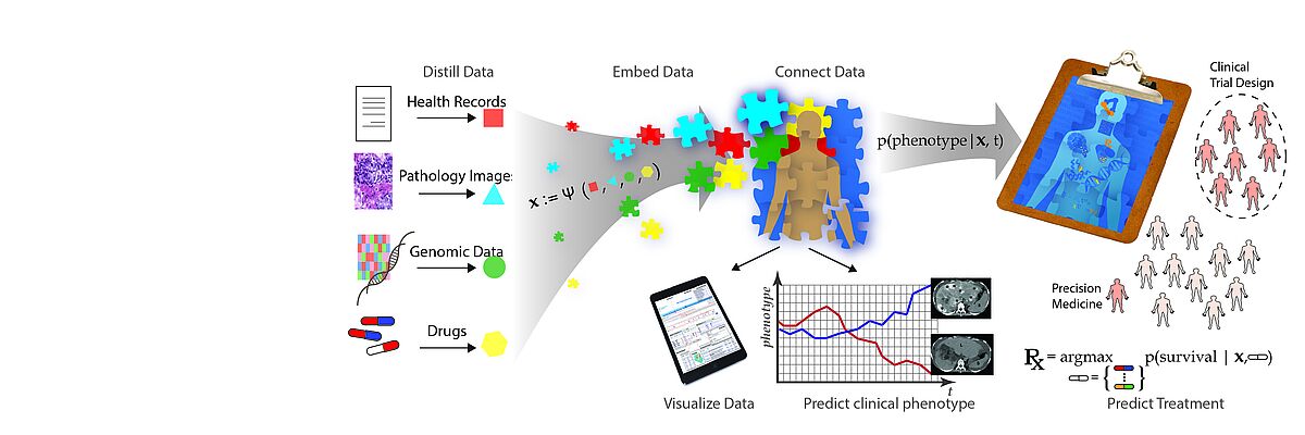
The emergence of data-driven medicine leverages data and algorithms to shape how we diagnose and treat patients. Machine Learning approaches allow us to capitalise on the vast amount of data produced in clinical settings to generate novel biomedical insights and build more precise predictive models of disease outcomes and treatment efficacy.
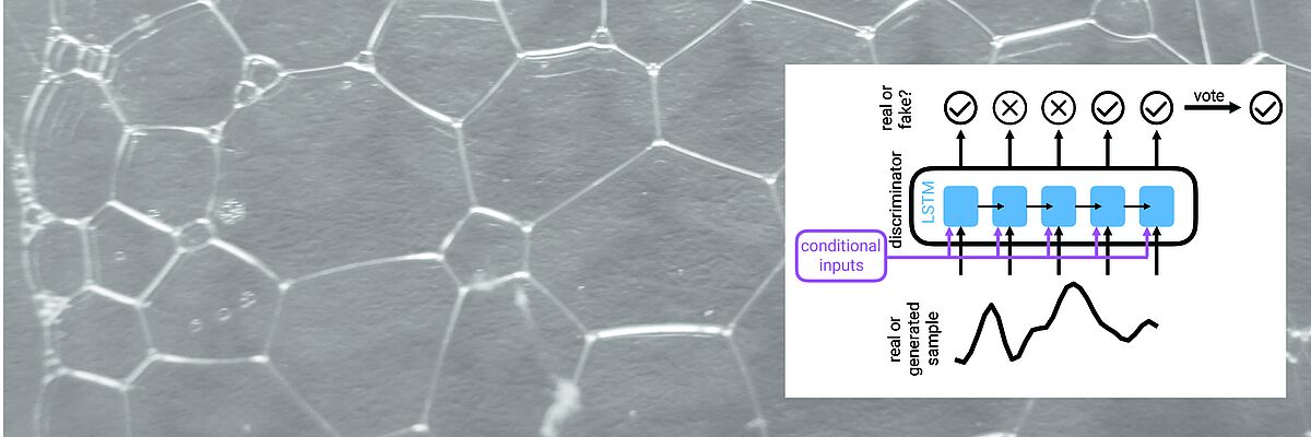
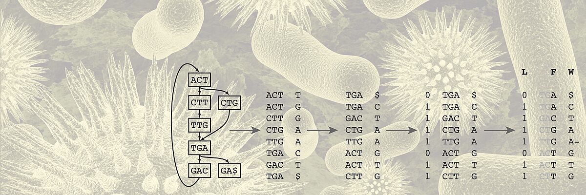
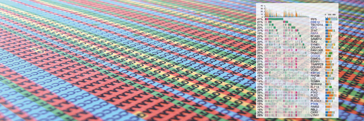
We work towards this transformation mainly but not exclusively in two key areas. One key application area is the analysis of heterogeneous data of cancer patients. For Genomics, we develop algorithms for storing, compressing, and searching extensive genomics datasets. Another key area is the development of time series models of patient health states and early warning systems for intensive care units.
Publications
Abstract Deep learning has enabled virtual spatial transcriptomics from histology, offering a scalable approach to molecular profiling of routinely archived tissue without molecular assays. However, reliable uncertainty estimates are essential for identifying potentially inaccurate predictions, and it remains unclear whether existing methods provide useful information about prediction reliability. Here, we present a systematic benchmark of uncertainty estimation for virtual spatial transcriptomics across three cancer cohorts spanning melanoma, lung, and kidney tumors. We evaluate aleatoric and epistemic uncertainty using probabilistic likelihoods and approximate Bayesian methods alongside histology-free and training-free baselines, assessing both calibration and error informativeness. Across cohorts and methods, uncertainty is associated with prediction error, but the predicted mean largely explains this association. After accounting for the mean, uncertainty provides only weak information about larger errors. Moreover, good calibration does not imply informative uncertainty, with simple distributions fitted without histology achieving comparable calibration. We therefore distinguish calibrated from informative uncertainty and argue that uncertainty should provide information about prediction error beyond the predicted mean. This benchmark provides a framework for systematic uncertainty evaluation in virtual spatial transcriptomics.
Authors Thomas Batique, Kalin Nonchev, Viktor Hendrik Koelzer, Gunnar Rätsch
Submitted NeurIPS 2026 ICBINB-BIO
Abstract Spatial transcriptomics pairs spatially resolved gene expression with tissue morphology in the same tissue section. Transcriptomic foundation models encode such expression profiles into general-purpose representations, but these representations can retain slide- and cohort-specific variation that obscures biological signal. Here, we test whether matched H&E histology can improve these representations as a training-time supervisory signal that is discarded at inference. Across three transcriptomic foundation models, histology-guided supervision provides no consistent aggregate improvement in cross-donor annotation transfer, despite substantially stronger transfer from histology alone. We find that gene-expression embeddings from spot-based spatial transcriptomics are low-dimensional and strongly structured by slide identity, limiting the shared gene-morphology signal available for cross-modal transfer. Guidance benefits some morphologically distinctive classes but reduces held-out gene predictivity broadly across the transcriptome. These results suggest that cross-modal supervision can reorganize information already encoded in a frozen representation but cannot recover information the representation does not retain, highlighting the importance of diagnosing slide-specific structure before applying such supervision.
Authors Ulysse McConnell, Kalin Nonchev, Viktor Hendrik Koelzer, Gunnar Rätsch
Submitted NeurIPS 2026 ICBINB-BIO
Abstract Motivation: Record-seq captures cumulative transcriptional activity over time in engineered Escherichia coli by integrating cellular RNA-derived spacer sequences into clustered regularly interspaced short palindromic repeats (CRISPR) arrays, which are read out by sequencing. Unlike the approximately uniform transcript sampling of RNA-seq, Record-seq records biological signal as spacers sampled by the CRISPR spacer acquisition machinery. Consequently, standard RNA-seq analysis strategies are not directly applicable, limiting sensitivity and interpretability. Our previous pipeline addressed these challenges only partially, retained inherited RNA-seq assumptions, and had limited algorithmic efficiency. Results: Here, we present an end-to-end computational framework for Record-seq data. To address the primary computational bottleneck of spacer sequence extraction, we implemented a wavefront alignment approach for efficient quasi-local pattern matching, achieving an approximately 30-fold speedup. We introduce transcription unit-based feature counting as an alternative to gene-body quantification to better represent prokaryotic transcription and increase statistical power by capturing signal from untranslated regions, which are spacer acquisition hotspots. For downstream analyses, we incorporate multiple normalization strategies and a nonparametric differential expression testing framework designed for sparse datasets. Further, we analyze spacer acquisition patterns and train sequence-based neural models that predict acquisition propensity from genomic sequence and annotations, providing a framework for assessing whether acquisition rules generalize as Record-seq is extended to new microbial hosts.
Authors Florian Hugi, Tanmay Tanna, Randall J. Platt
Submitted Bioinformatics
Abstract Spatial transcriptomics remains costly and low-throughput, limiting it to a small fraction of routine histology and leaving the molecular state of disease unmeasured in most patients. Predicting spatial expression from histology could address this gap, but existing methods are restricted to predefined genes and small cohorts. We present DeepSpot-M, a multimodal foundation model that predicts spatial expression by representing genes with embeddings from foundation models spanning DNA, RNA, proteins, single cells and biomedical text. By reformulating prediction as a query over genes, DeepSpot-M spans the protein-coding transcriptome and predicts genes unseen during training. Trained on a large pan-cancer dataset, it transfers to held-out cancers, outperforming specialised models trained on them, and adapts to new cohorts and single-cell assays from one slide via test-time adaptation. Applied to TCGA, it generates a virtual atlas of 28,664 slides across 32 cancers, recovering a pan-cancer map of malignancy from histology. The same query interface further enables transcriptome restoration, cross-species non-coding RNA inference, in silico variant-effect mapping and natural-language querying. We anticipate DeepSpot-M will provide a scalable foundation for virtual spatial transcriptomics and biomarker discovery.
Authors Kalin Nonchev, Sebastian Dawo, Karina Silina, Viktor H Koelzer, Gunnar Rätsch
Submitted medRxiv
Abstract Spatial transcriptomics couples hematoxylin and eosin (H&E) tissue morphology with spatially resolved gene expression (GE). However, generative models that exploit this coupling to synthesize tissue images from transcriptomic profiles remain scarce. We present STMDiT (Spatial Transcriptomics and Morphology Diffusion Transformer), a diffusion transformer that synthesizes H&E histopathology patches conditioned jointly on morphological embeddings and transcriptomic profiles. Building on PixCell (Yellapragada et al., 2025), we integrate gene expression from a frozen CancerFoundation encoder (Theus et al., 2024) through adaptive layer normalization and per-block cross-attention, and we train under dual classifier-free guidance with independent modality dropout. On the 10x TuPro Visium melanoma cohort, GE conditioning improves both image quality over the no-GE PixCell-B baseline (best FID = 252.9 vs 330.7) and transcriptomic fidelity (best AUC = 0.267 vs 0.229, reaching 82% of the real-tile ceiling). Training with DeepSpot's predicted-transcriptomics pseudo-labels (PTPL) uniquely transfers zero-shot to TCGA SKCM, an out-of-distribution (OOD) H&E-only melanoma cohort: PTPL-XAttn-PMA-B reaches FID = 690.0, a 57-point improvement over the no-GE baseline (747.1), with a within-model GE-ablation effect of ΔOOD = +309.5, enabling virtual tissue synthesis beyond native spatial-transcriptomics coverage. Our results indicate that gene-expression conditioning produces morphologically distinct tissue images and supports virtual tissue simulation for hypothesis testing in computational pathology. Code availability: https://github.com/ratschlab/stmdit
Authors Pantelis Vlachas, Kalin Nonchev, Viktor H Koelzer, Gunnar Rätsch
Submitted ICML 2026 Multi-modal Foundation Models and Large Language Models for Life Sciences


