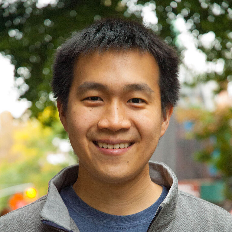
David Kuo,

"Nothing in Biology Makes Sense Except in the Light of Evolution" - Dobzhansky (1973)
Alumni
- Address
-
MSKCC – Computational Biology Center
1275 York Avenue, Box # 357
New York, NY 10065 - Room
- MSKCC: Z-677, HSS: 8th Floor
- @dkuo
I studied Biology at Stanford University and worked as a Developer at McMaster-Carr Supply
Company before beginning my PhD studies in Systems Biology at Weill Cornell Medicine in New York City.
I began biomedical research in the laboratory of alternative splicing expert Jane Wu in the summers from 2001-2005. I earned my BS in Biological Sciences at Stanford University in 2008. While at Stanford, I performed research in the laboratory of cancer biologist Calvin Kuo (no relation), where we studied the effects of VEGF blockade on liver vasculature. After college, I worked as an Information Systems Developer for McMaster-Carr Supply Company before matriculating at the Weill Cornell Graduate School of Medical Sciences in the Physiology, Biophysics & Systems Biology Department. I joined the Rätsch Lab in 2012 with primary interests in genomics and biological data analysis. I collaborate actively with the laboratories of Lionel Ivashkiv at Hospital for Special Surgery and the Hans-Guido Wendel at MSKCC.
Latest Publications
Abstract The activation of memory T cells is a very rapid and concerted cellular response that requires coordination between cellular processes in different compartments and on different time scales. In this study, we use ribosome profiling and deep RNA sequencing to define the acute mRNA translation changes in CD8 memory T cells following initial activation events. We find that initial translation enables subsequent events of human and mouse T cell activation and expansion. Briefly, early events in the activation of Ag-experienced CD8 T cells are insensitive to transcriptional blockade with actinomycin D, and instead depend on the translation of pre-existing mRNAs and are blocked by cycloheximide. Ribosome profiling identifies ∼92 mRNAs that are recruited into ribosomes following CD8 T cell stimulation. These mRNAs typically have structured GC and pyrimidine-rich 5′ untranslated regions and they encode key regulators of T cell activation and proliferation such as Notch1, Ifngr1, Il2rb, and serine metabolism enzymes Psat1 and Shmt2 (serine hydroxymethyltransferase 2), as well as translation factors eEF1a1 (eukaryotic elongation factor α1) and eEF2 (eukaryotic elongation factor 2). The increased production of receptors of IL-2 and IFN-γ precedes the activation of gene expression and augments cellular signals and T cell activation. Taken together, we identify an early RNA translation program that acts in a feed-forward manner to enable the rapid and dramatic process of CD8 memory T cell expansion and activation.
Authors Darin Salloum, Kamini Singh, Natalie R Davidson, Linlin Cao, David Kuo, Viraj R Sanghvi, Man Jiang, Maria Tello Lafoz, Agnes Viale, Gunnar Ratsch, Hans-Guido Wendel
Submitted The Journal of Immunology
Abstract Macrophages tailor their function according to the signals found in tissue microenvironments, assuming a wide spectrum of phenotypes. A detailed understanding of macrophage phenotypes in human tissues is limited. Using single-cell RNA sequencing, we defined distinct macrophage subsets in the joints of patients with the autoimmune disease rheumatoid arthritis (RA), which affects ~1% of the population. The subset we refer to as HBEGF+ inflammatory macrophages is enriched in RA tissues and is shaped by resident fibroblasts and the cytokine tumor necrosis factor (TNF). These macrophages promoted fibroblast invasiveness in an epidermal growth factor receptor–dependent manner, indicating that intercellular cross-talk in this inflamed setting reshapes both cell types and contributes to fibroblast-mediated joint destruction. In an ex vivo synovial tissue assay, most medications used to treat RA patients targeted HBEGF+ inflammatory macrophages; however, in some cases, medication redirected them into a state that is not expected to resolve inflammation. These data highlight how advances in our understanding of chronically inflamed human tissues and the effects of medications therein can be achieved by studies on local macrophage phenotypes and intercellular interactions.
Authors David Kuo, Jennifer Ding, Ian Cohn, Fan Zhang, Kevin Wei, Deepak Rao, Cristina Rozo, Upneet K Sokhi, Sara Shanaj, David J. Oliver, Adriana P. Echeverria, Edward F. DiCarlo, Michael B. Brenner, Vivian P. Bykerk, Susan M. Goodman, Soumya Raychaudhuri, Gunnar Rätsch, Lionel B. Ivashkiv, Laura T. Donlin
Submitted Science Translational Medicine
Abstract Macrophages tailor their function to the signals found in tissue microenvironments, taking on a wide spectrum of phenotypes. In human tissues, a detailed understanding of macrophage phenotypes is limited. Using single-cell RNA-sequencing, we define distinct macrophage subsets in the joints of patients with the autoimmune disease rheumatoid arthritis (RA), which affects ~1% of the population. The subset we refer to as HBEGF+ inflammatory macrophages is enriched in RA tissues and shaped by resident fibroblasts and the cytokine TNF. These macrophages promote fibroblast invasiveness in an EGF receptor dependent manner, indicating that inflammatory intercellular crosstalk reshapes both cell types and contributes to fibroblast-mediated joint destruction. In an ex vivo tissue assay, the HBEGF+ inflammatory macrophage is targeted by several anti-inflammatory RA medications, however, COX inhibition redirects it towards a different inflammatory phenotype that is also expected to perpetuate pathology. These data highlight advances in understanding the pathophysiology and drug mechanisms in chronic inflammatory disorders can be achieved by focusing on macrophage phenotypes in the context of complex interactions in human tissues.
Authors David Kuo, Jennifer Ding, Ian Cohn, Fan Zhang, Kevin Wei, Deepak Rao, Cristina Rozo, Upneet K Sokhi, Accelerating Medicines Partnership RA/SLE Network, Edward F. DiCarlo, Michael B. Brenner, Vivian P. Bykerk, VSusan M. Goodman, Soumya Raychaudhuri, Gunnar Rätsch, Lionel B. Ivashkiv, Laura T. Donlin
Submitted bioRxiv
Abstract Insulin initiates diverse hepatic metabolic responses, including gluconeogenic suppression and induction of glycogen synthesis and lipogenesis. The liver possesses a rich sinusoidal capillary network with a higher degree of hypoxia and lower gluconeogenesis in the perivenous zone as compared to the rest of the organ. Here, we show that diverse vascular endothelial growth factor (VEGF) inhibitors improved glucose tolerance in nondiabetic C57BL/6 and diabetic db/db mice, potentiating hepatic insulin signaling with lower gluconeogenic gene expression, higher glycogen storage and suppressed hepatic glucose production. VEGF inhibition induced hepatic hypoxia through sinusoidal vascular regression and sensitized liver insulin signaling through hypoxia-inducible factor-2α (Hif-2α, encoded by Epas1) stabilization. Notably, liver-specific constitutive activation of HIF-2α, but not HIF-1α, was sufficient to augment hepatic insulin signaling through direct and indirect induction of insulin receptor substrate-2 (Irs2), an essential insulin receptor adaptor protein. Further, liver Irs2 was both necessary and sufficient to mediate Hif-2α and Vegf inhibition effects on glucose tolerance and hepatic insulin signaling. These results demonstrate an unsuspected intersection between Hif-2α-mediated hypoxic signaling and hepatic insulin action through Irs2 induction, which can be co-opted by Vegf inhibitors to modulate glucose metabolism. These studies also indicate distinct roles in hepatic metabolism for Hif-1α, which promotes glycolysis, and Hif-2α, which suppresses gluconeogenesis, and suggest new treatment approaches for type 2 diabetes mellitus.
Authors K Wei, SM Piecewicz, LM McGinnis, CM Taniguchi, SJ Wiegand, K Anderson, CW M Chan, KX Mulligan, David Kuo, J Yuan, M Vallon, LC Morton, E Lefai, MC Simon, JJ Maher, G Mithieux, F Rajas, JP Annes, OP McGuinness, G Thurston, AJ Giaccia, CJ Kuo
Submitted Nat Med